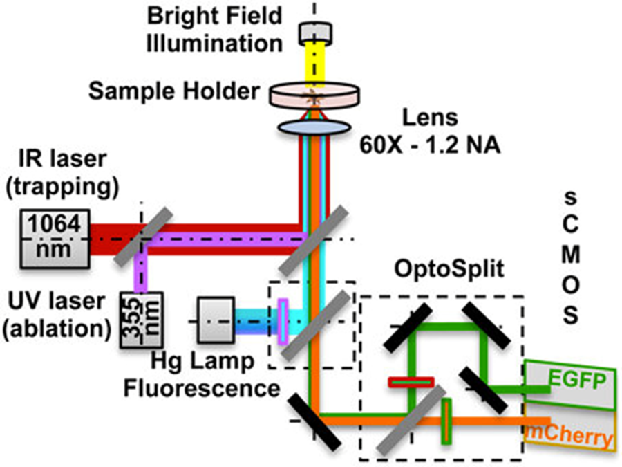
Unveiling the Cellular Orchestra: Advanced Imaging Techniques for Multiple Signals
Cells are like tiny orchestras, with a complex symphony of signaling molecules dictating their every move. Understanding these intricate communication pathways is crucial for unlocking the mysteries of health, disease, and development. But traditional imaging techniques often struggle to capture the full picture, limited in the number of signals they can detect simultaneously.
This blog post delves into the world of advanced imaging techniques that are revolutionizing our ability to visualize multiple cellular signals. We’ll explore how these methods work, their advantages, and the exciting new insights they’re providing into cellular function.
The Challenges of Multi-Signal Imaging
Before diving into the exciting world of advanced techniques, let’s acknowledge the limitations of conventional methods:
Limited Multiplexing: Traditional techniques like fluorescence microscopy can only visualize a handful of signals simultaneously, often requiring separate experiments for each marker.
Resolution vs. Multiplexing: Techniques offering higher resolution often sacrifice the ability to track multiple signals.
Specificity: Differentiating between different fluorescent labels can be challenging, leading to signal overlap and inaccurate data.
Standard microscopy techniques, like fluorescence microscopy, use fluorescent tags to visualize specific molecules within a cell. However, they are typically limited to detecting a few different signals at once. This is because the fluorescent tags all emit light at similar wavelengths, making it difficult to distinguish between them.
The Power of Multiplexed Imaging
Multiplexed imaging techniques overcome this limitation by employing various strategies to differentiate between multiple signals. Here are some of the leading contenders:
Multiplexed Fluorescence Microscopy with Switchable Fluorophores
This technique utilizes a clever strategy. Scientists have engineered fluorescent molecules that can be toggled “on” and “off” at specific rates. Each molecule emits a distinct color (red or green) and blinks at a different frequency. By labeling cellular components of interest with these switchable fluorophores, researchers can image the cell over time. Clever computational analysis, similar to how our ears distinguish musical notes, separates the blinking signals, revealing the location and activity of each labeled molecule within the cell.
This method offers several advantages:
- Wide Applicability: It can be implemented on standard microscopes, making it accessible to many labs.
- High Multiplexing: The ability to use multiple fluorophores with distinct blinking rates allows for visualizing up to seven different signals simultaneously, with the potential for even more in the future.
- Live Cell Imaging: This technique can be used to study dynamic cellular processes in real-time.
Spectral Imaging Techniques
These methods leverage the entire spectrum of light emitted by fluorescent molecules to differentiate between them. By analyzing the unique spectral signature of each fluorophore, researchers can distinguish closely related fluorescent labels, enabling the visualization of more signals simultaneously.
FCS (Fluorescence Correlation Spectroscopy) and RICS (Raster Image Correlation Spectroscopy)
These techniques measure the diffusion and interaction of fluorescent molecules within a cell. By analyzing the fluctuations in fluorescence intensity, researchers can gain insights into molecular dynamics and binding events. While not directly providing spatial information, FCS and RICS offer valuable information on cellular signaling interactions.
Super-resolution Microscopy
This category of techniques pushes the boundaries of resolution, allowing researchers to visualize cellular structures and molecules at a much finer scale than traditional methods. Some popular examples include:
STORM (Stochastic Optical Reconstruction Microscopy) and PALM (Photoactivated Localization Microscopy)
These techniques achieve super-resolution by precisely localizing individual fluorescent molecules within a cell. This allows for the visualization of closely spaced proteins and organelles that would be indistinguishable with standard microscopy.
SIM (Structured Illumination Microscopy)
This approach utilizes patterned light to create a high-resolution image, offering improved resolution compared to conventional fluorescence microscopy.
While super-resolution techniques offer fantastic detail, they typically struggle with multiplexing. However, ongoing research is exploring ways to combine super-resolution with multiplexing capabilities.
Fluorescence Lifetime Imaging Microscopy (FLIM)
This technique measures the time it takes for a fluorescent molecule to return to its ground state after being excited by light. Different fluorophores have unique lifetimes, allowing researchers to distinguish them even if they emit light at similar wavelengths.
Förster Resonance Energy Transfer (FRET)
FRET utilizes the non-radiative transfer of energy between two fluorescent molecules in proximity. By carefully selecting donor and acceptor fluorophores, scientists can monitor protein-protein interactions within a cell.
Cyclic Fluorophore Activation and Deactivation (cfPAINT)
This innovative approach uses fluorescent tags that can be switched on and off with light. By employing different blinking patterns for various tags, researchers can track the abundance and localization of multiple molecules simultaneously.
Benefits and Applications
Multiplexed imaging offers a plethora of advantages
Unveiling Complex Signaling Networks
By visualizing multiple signals at once, scientists can gain a deeper understanding of how different pathways interact and influence cellular behavior.
Improved Diagnosis and Drug Discovery
These techniques can aid in identifying cellular abnormalities associated with diseases, leading to more targeted therapies.
Studying Dynamic Processes
Multiplexed imaging allows researchers to track changes in cellular signalling over time, providing insights into dynamic processes like cell division and differentiation.
The Future of Multi-Signal Imaging
The field of advanced imaging is rapidly evolving. Here’s a glimpse into what the future holds
Further Development of Multiplexing Techniques
Researchers are constantly innovating new methods for labelling and differentiating multiple signals within a cell. Expect even higher multiplexing capabilities in the years to come.
Integration with Artificial Intelligence (AI)
AI algorithms are becoming increasingly powerful in analyzing complex image data. Integrating AI with advanced imaging techniques will facilitate faster and more accurate data interpretation.
Live-Cell Imaging Advancements
The ability to study dynamic cellular processes in real-time is crucial for understanding cellular signaling. Expect further advancements in techniques that enable long-term, high-resolution live-cell imaging of multiple signals.
Featured Image:
Source:

